
The goals of this experiment are to learn the operating principles of a scanning tunneling microscope (STM), accurately calibrate the STM by imaging a gold grating, and to study the surface of graphite (HOPG).
|
|
The goals of this experiment are to learn the operating principles of a scanning tunneling microscope (STM), accurately calibrate the STM by imaging a gold grating, and to study the surface of graphite (HOPG). |
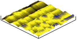 |
The experiment begins with calibration of the STM. This is done by imaging a gold grating with known grating spacing. The scanning range of the STM is then compared to the size of the scanned image (determined from known grating spacing). This comparison allows the calculation of a scale factor, so that horizontal dimensions on the sample can be accurately determined. |
This is an image of an Au grating. (Image by Robert Carver, James Little, Fall 1997) |
|
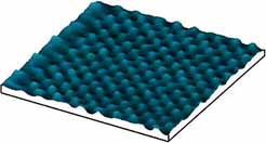
The following step is to scan HOPG (Highly Oriented Pyrolitic Graphite). These images should reveal the structure of Carbon atoms within HOPG. Bond lengths and angles can be determined using the HOPG images and the scan range of the image, determined from the calibrated scale factor. |
|
This is a 3 dimensional, 20x20 nm image of HOPG. The mounds correspond to Carbon atoms. (Image by Matt Cowan, Fall 1997) |
Senior Lab Home |